Original Title: Transfemoral Access Assessment for Transcatheter Aortic Valve Replacement Evidence Based Application of Computed Tomography over Invasive Angiography. Reference: Circ Cardiovas Imaging 2015 DOI:10.1161/CIRCIMAGING.114.0011995.
Courtesy of Dr. Guillermo Migliaro
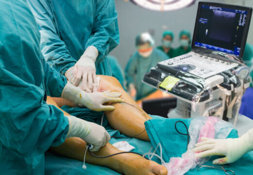
Transfemoral access for TAVI was introduced in 2006 and is considered first choice. Vascular complications (VCs) are important events that correlate to increased mortality. According to the Valve Academic Research Consortium I (VARC) major VCs are observed in 5 to 23.3% of patients and minor VCs, in 5.6 to 28.3%.
Access site assessment is crucial to prevent post TAVI vascular complications. Vessel diameter, calcification and tortuosity in iliofemoral access vessels are important values that determine the risk of sheath insertion related vascular complications. Among these, vessel diameter in relation to sheath size is considered the factor that best predicts vascular complications. Even though the CT is commonly used to evaluate access sites at iliac and femoral level, no study to date has assessed the value of measurements derived from CT over angiography for the prediction of post TAVI VCs.
The purpose of this study was to compare the predictive value of CT over angiography, to prevent vascular complications and identify the imaging strategy that best predicts vascular complications.
The study reviewed retrospective data from 486 consecutive transfemoral TAVI patients that had received the balloon expandable Edward Sapien in one single center. Primary end point was a combination of sheath related complications defined as iliofemoral arterial injury caused by catheter manipulation (excluding VC related to cannulation site, since these could be caused by multiple factors). Receiver operating characteristic models (ROC) were generated using sheath-to-iliofemoral artery ratios as a variable and SRC as an end point.
2 cohorts were generated: a contrast CT cohort in which patients had both contrast CT and angiography (n=283), and another in which patients had non contrast CT and angiography (n=103). 110 patients that had only one of the methods were excluded from the analysis.
In patients undergoing contrast CT and angiography (n=283; 35 SRCs), contrast CT showed a greater predictive value than angiography by area under the curve (p<0.001): 0.87 (95% CI 0.82–0.91) versus 0.72 (95% CI 0.66–0.77). In patients undergoing non contrast CT and angiography (n=103; 17 SRCs), there were no significant differences in areas under the curve 0.79 (95% CI 0.70–0.86) versus 0.73 (95% CI 0.63–0.81). Assessments of calcification and tortuosity provided no useful value for SRC prediction.
Conclusion
Assessing vessel diameter by contrast CT has a greater predictive value for post-transcatheter aortic valve replacement vascular complications than angiography, which is why TAVI access site should be evaluated by contrast CT whenever possible.
Editorial Comment
This is a retrospective study, not randomized, carried out in one single center that presents low events incidence and uses only one valve, unavailable in many countries in Latin America. As in the case of valve annulus measurement, CT seems to be the best method to assess TAVI access site and prevent vascular complications.