

Despite robust information supporting the use of intravascular imaging during coronary angioplasty, its use in clinical practice remains low. This paper proposes using an algorithm for decision-making throughout the procedure to promote increased intravascular imaging use.
Regardless of the many technologies that have been incorporated into strut design struts and polymer type, the rate of stent failure has not changed significantly in recent years.
Outcome improvement seems not to depend on a new and better stent, but on a refined implantation technique. Here is where intravascular imaging—which should already be standard of care—becomes essential.
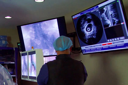
In spite of their significant differences, intravascular ultrasound (IVUS) and optical coherence tomography (OCT) can be used interchangeably in most clinical scenarios. Both technologies allow co-registration with angiography and functional measurements.
Read also: Risk of Coronary Obstruction in Repeat TAVR.
However, there are some specific cases where one technology is clearly more beneficial than the other.
- Left main coronary artery disease: IVUS is better than OCT in evidence and practicality, allowing the evaluation of both the bifurcation and the ostium.
- Ostial lesion: IVUS is the clear choice. The need to inject contrast to have clear images with OCT is hampered by not being able to intubate the guidewire.
- In-stent restenosis: Both technologies are useful, but OCT comes on top because of its much sharper images to define malapposition or lack of strut coverage.
- Calcium: Both tools are suitable; however, OCT is preferred—again—because of its high resolution.
- Chronic total occlusion: IVUS is more useful to direct the guidewire to the true lumen.
- Stent sizing and optimization: Both technologies are valuable. The minimal in-stent lumen area is the greatest predictor of events, and it can be measured and optimized with both tools.
- Plaque rupture and thrombosis: In acute coronary syndromes, OCT is a clear winner in finding the culprit lesion. OCT advantages include the possibility of observing plaque rupture, measuring cap thickness, detecting inflammation, and more.
- Chronic kidney disease: The need to inject contrast and the limitation in the length of the pullbacks make OCT unsuitable for patients with renal impairment.
Regardless of their differences (and their availability), using at least one of them is paramount. Currently, intravascular imaging is the best hope for improving angioplasty results.
Original title: Intravascular Imaging-Guided Percutaneous Coronary Intervention. A Universal Approach for Optimization of Stent Implantation.
Reference: Evan Shlofmitz, DO et al. Circ Cardiovasc Interv. 2020;13:e008686. DOI: 10.1161/CIRCINTERVENTIONS.120.008686.
Subscribe to our weekly newsletter
Get the latest scientific articles on interventional cardiology


