

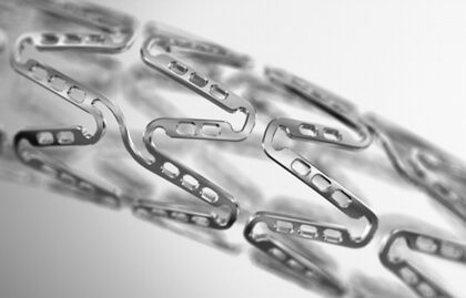
A year after the implantation of everolimus-eluting bioresorbable scaffold Absorb 1.1, optical coherence tomography (OCT) scans showed neointima formation covering plaque without significant luminal loss. At 5 years, the device is supposed to disappear completely, potentially thrombogenic plaque components are supposed to be covered by endothelium, and the vasomotor function is supposed to be recovered.
 However, there are reports on scaffold persistence at 5 years and little information on final artery healing as regards neointimal tissue and eventual neoatherosclerosis, both in and out of the treated segment.
This study included patients who had taken part in the ABSORB EXTEND trial and who underwent OCT imaging at baseline after the index procedure and at 1 and 5 years. Plaque distributions both in and out of treated segments were analyzed. Among patients enrolled, 25% had diabetes and were stable at the time the index procedure was carried out.
Read also: Leaflet Laceration, an Extreme Measure to Avoid Coronary Occlusion After TAVR.
At 5 years, compared with baseline imaging from segments treated with bioresorbable scaffolds at the time of the index procedure, there was a higher prevalence of lipid-laden neointima (17% vs. 61%; p = 0.04); higher calcification (28% vs. 94%; p < 0.01); higher neovasculatization (6% vs. 78%; p < 0.01), and more thin-cap fibroatheroma (0% vs. 22%; p = 0.02).
Such plaque progression was not observed in untreated segments at 1 and 5 years.
Conclusion
This small study showed the occurrence and progression of neoatherosclerosis with luminal narrowing at 5 years in segments treated with first-generation bioresorbable scaffolds. Since initial expectations regarding this new technology had to do precisely with their long-term benefits, these findings warrant confirmation in larger studies.
Original title: Neoatherosclerosis 5 Years After Bioresorbable Vascular Scaffold Implantation.
Reference: Noriaki Moriyama et al. J Am Coll Cardiol 2018;71:1882-93.
Get the latest scientific articles on interventional cardiologySubscribe to our weekly newsletter
We are interested in your opinion. Please, leave your comments, thoughts, questions, etc., below. They will be most welcome.


