Computerized Tomography as First Coronary Study?

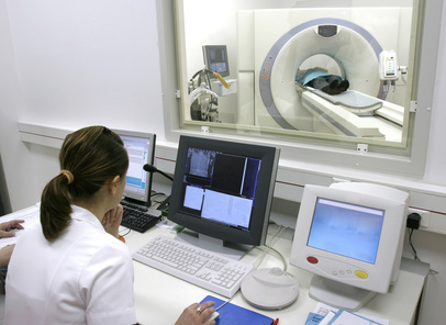
According to this new document recently published in JACC, a computerized tomography (CT) coronary angiography should be the first study done to diagnose a stable coronary artery disease.

ffr tomografia

A multidisciplinary group of experts recommends using computerized tomography as first-line diagnosis in stable patients. According to these professionals, guidelines should be updated soon.

Given the accruing evidence favoring anatomical studies (such as the PROMISE, the SCOT-HEART, and, more recently, the ISCHEMIA), this expert committee believes that computerized tomography offers the greatest benefit for patient management.

Despite the evidence supporting the benefits and even the savings in patients without known coronary disease, reality indicates that, for every 60 gamma-camera studies (single photon emission computed tomography, SPECT) only one tomography is conducted.


Read also: Visual Assessment for Non-Culprit Lesion Revascularization.


his could be partially due to the vast number of centers with nuclear medicine and stress echocardiography, the traditional idea that function supersedes anatomy, and the administrative difficulties faced by health systems in the payment of tomographies.

Both United Kingdom and European guidelines have been updated with the use of CT in chronic coronary syndromes, but the old school approach is still going strong in the United States. There might also be a lack of trained staff who can interpret the images.

We might be reaching a time when computerized tomography scans will completely replace us, and we will only enter the cath lab for therapeutic procedures.

Original Title: Current evidence and recommendations for coronary CTA first in evaluation of stable coronary artery disease.

Reference: Poon M et al. J Am Coll Cardiol. 2020;76:1358-1362.


Subscribe to our weekly newsletter

Get the latest scientific articles on interventional cardiology

We are interested in your opinion. Please, leave your comments, thoughts, questions, etc., below. They will be most welcome.

More articles by this author

Plaque Ruptures in Non-Culprit Arteries: Follow-Up With Intravascular Imaging

Plaque rupture remains one of the most important pathophysiological mechanisms in acute coronary syndromes. However, not all ruptures manifest clinically as ischemia, myocardial infarction,...

OCT-Detected High-Risk Plaques Predict Recurrent Events After Myocardial Infarction

After a myocardial infarction (MI), non-culprit lesions are often deferred when they are not flow-limiting (negative FFR). However, these lesions continue to represent an...

Ticagrelor vs Clopidogrel in ACS Patients Receiving DOAC After PCI: More Bleeding Without Ischemic Benefit?

In patients with acute coronary syndrome (ACS) who require direct oral anticoagulation (DOAC) and undergo percutaneous coronary intervention (PCI), current guidelines recommend a dual...

EuroPCR 2026 | 10-Year Left Main PCI: When Survival Is Similar, Should the Less Invasive Strategy Prevail?

The primary goal of revascularization in left main coronary artery disease (LMCA) is to improve survival. However, debate continues regarding whether, in anatomically suitable...

LEAVE A REPLY

Please enter your comment!
Please enter your name here

Related Articles

SOLACI Sessionsspot_img
Jornadas Guatemala 2026

Recent Articles

Plaque Ruptures in Non-Culprit Arteries: Follow-Up With Intravascular Imaging

Plaque rupture remains one of the most important pathophysiological mechanisms in acute coronary syndromes. However, not all ruptures manifest clinically as ischemia, myocardial infarction,...

Drug-Eluting Stents in Peripheral Arterial Disease: When Should They Be Used?

Peripheral drug-eluting stents have transformed the treatment of peripheral arterial disease by reducing restenosis rates and the need for repeat interventions. However, the emergence...

OCT-Detected High-Risk Plaques Predict Recurrent Events After Myocardial Infarction

After a myocardial infarction (MI), non-culprit lesions are often deferred when they are not flow-limiting (negative FFR). However, these lesions continue to represent an...