

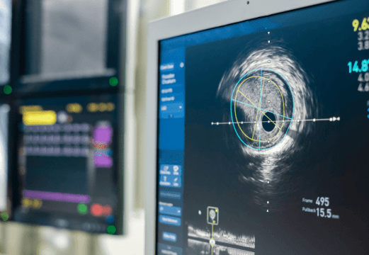
Plaque rupture remains one of the most important pathophysiological mechanisms in acute coronary syndromes. However, not all ruptures manifest clinically as ischemia, myocardial infarction, or angina. In the absence of thrombus, or when thrombus is insufficient to compromise coronary flow, these disruptions may remain clinically silent. In this context, the characterization of non-obstructive lesions in non–infarct-related arteries (non-IRA) becomes particularly relevant, especially through the use of high-resolution intracoronary imaging modalities such as OCT, IVUS, and NIRS.
The aim of the study was to evaluate the frequency and characteristics of silent plaque ruptures in non-obstructive non-IRA lesions, analyze their morphological evolution at 52 weeks, and identify the morphology of lesions that developed new ruptures during follow-up.
Data from the IBIS-4 and PACMAN-AMI studies were analyzed. Both included patients with myocardial infarction successfully treated with PCI of the culprit lesion. The analysis included 336 patients, 613 non-IRAs, and 783 lesions defined by IVUS. The evaluated lesions were non-obstructive, with stenosis <50%. Plaque rupture was defined by OCT as a discontinuity of the intimal layer with communication between the cavity and the coronary lumen. Healing was defined as the disappearance of the flap and cavity at the rupture site.
Plaque rupture was identified in 41 lesions from 40 patients, representing 12% of patients and 5% of the analyzed lesions. Multiple rupture sites were observed in 5 lesions, and thrombus was documented in 10 of the 41 lesions with rupture, predominantly red thrombus according to OCT findings.
From a morphological standpoint, lesions with rupture exhibited a more vulnerable phenotype, with a higher percentage atheroma volume by IVUS (53.3±6.4% vs 49.5±5.8%; 95% CI 1.9–5.4; p<0.001) and a lower minimum fibrous cap thickness by OCT (70±49 vs 116±84 μm; 95% CI -75 to -11; p=0.009).
Read also: Drug-Eluting Stents in Peripheral Arterial Disease: When Should They Be Used?
In the multivariate analysis, percentage atheroma volume was independently associated with plaque rupture (OR 1.11 per 1% increase; 95% CI 1.05–1.18; p<0.001), as were mean external elastic membrane area (OR 1.17 per 1 mm² increase; 95% CI 1.10–1.24; p<0.001) and the presence of fibroatheroma (OR 2.46; 95% CI 1.16–5.48; p=0.022).
During scheduled follow-up, among the 41 rupture sites assessed by OCT, 51% showed healing at 52 weeks. Final morphology was predominantly fibrous plaque, followed by layered plaque and thick-cap fibroatheroma. Evaluation of lesion evolution identified 10 new silent ruptures, more frequently observed in lesions with thin-cap fibroatheroma morphology, followed by thick-cap fibroatheroma, layered plaque, and hematoma.
Read also: OCT-Detected High-Risk Plaques Predict Recurrent Events After Myocardial Infarction.
Clinical events at 1 year did not show an increased risk associated with silent rupture; the composite MACE endpoint occurred in 10% of patients with rupture and in 14% of those without rupture.
Conclusions: Silent Plaque Ruptures in Non-Culprit Arteries Show High Vulnerability and Stable Evolution at 1 Year
This study demonstrates that silent plaque ruptures in non-obstructive lesions of non–infarct-related arteries are relatively frequent in patients with myocardial infarction and are mainly associated with local vulnerability features. More than half of the ruptures evolved toward stable morphologies at 52 weeks, without showing a significant clinical association with cardiovascular events at 1 year.
Original Title: Silent plaque ruptures in non-obstructive lesions of non–infarct-related arteries: a multimodality, serial intracoronary imaging study.
Reference: Ryota Kakizaki, Flavio G Biccirè, Sylvain Losdat, Yasushi Ueki, Jonas D Häner, Hiroki Shibutani, Tatsuhiko Otsuka, Sarah Bär, Jacob Lønborg, Ernest Spitzer, George C M Siontis, Anna S Ondracek, Robert-Jan van Geuns, Yu-Jen Wang, Christian M Matter, Juan F Iglesias, David Spirk, Joost Daemen, Gregor Fahrni, Felix Mahfoud, Thomas Engstrøm, Irene M Lang, Konstantinos C Koskinas, Lorenz Räber. European Heart Journal, Volume 47, Issue 22, 7 June 2026, Pages 2814–2826. https://doi.org/10.1093/eurheartj/ehag037.


