

Predictors of intrastent restenosis in the right coronary artery ostium.
The ostium of the right coronary artery (RCA) presents certain histological aspects. Atherosclerotic and fibrotic plaques in this area contain an abundance of smooth muscle, collagen, and a certain degree of calcification, along with thicker adventitia. Additionally, it has certain anatomical aspects such as poor distensibility and excessive oscillating movement.
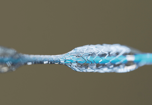
During the era of balloon angioplasty, the fact that lesions in this area had a lower success rate (88%) with a high percentage of acute complications was well-known. These events were mostly due to greater recoil compared with lesions in areas other than the right coronary artery. In the era of drug-eluting stents (DES), percutaneous coronary interventions (PCI) of the RCA ostium have shown a high prevalence of intrastent restenosis (ISR), reaching as high as 7.5-12.7%.
The aim of this study presented by Yamamoto K. et al. was to investigate the mechanisms of intrastent restenosis in the RCA ostium using intravascular ultrasound (IVUS). Researchers conducted an observational, retrospective, single-center study that included patients with intrastent restenosis (defined as stenosis ≥50% or a minimal luminal area ≤4 mm by IVUS) in the RCA ostium (within 3 mm from the aorto-ostial junction).
The primary endpoint (PFP) was a composite outcome called target lesion failure (TLF), which consisted of cardiac death, target vessel revascularization, and myocardial infarction of the treated vessel.
Read also: Frequency and Causes of Mortality in Chronic Total Occlusion.
A total of 139 lesions with ostial ISR were included. The mean age was 71 years, 42.4% of patients were women, 10.1% had aortic stenosis (at least moderate), and the majority of ISR lesions were focal.
The patterns of ISR were neointimal hyperplasia (25%), neoatherosclerosis (22%), uncovered ostial lesion (6%), stent underexpansion (11%), protrusion of a calcified nodule (11%), and stent fracture or deformation (25%).
The time interval between stent implantation and ISR was shorter in patients with uncovered ostiums (0.5 [0.2-0.6] years) compared with the interval in cases of neoatherosclerosis (2.7 [1.0-5.0] years). Stents were placed in 66.9% of treated lesions, and the stenting rate in ISR due to mechanical causes (49.3%) was lower compared with that in ISR due to biological causes (82.4%).
Read also: Minimal Stent Area: New IVUS Parameter?
The incidence of the primary endpoint at one year was 11.5%. When analyzing patients with a mechanical cause and no treatment with a new stent, they experienced a higher rate of TLF (41.4%) compared with other groups (P < 0.0001).
Conclusions
Approximately half of the cases of ISR were due to mechanical causes (stent fracture, stent underexpansion, or calcified protrusion). Subsequent events were more common in those patients, especially with mechanical causes and no new stent implantation.
Therefore, when treating this area, the use of an uncovered ostium and proper stent apposition should be avoided, favoring the use of IVUS to improve positioning. In cases of fracture, consideration should be given to using more robust stents to prevent such outcomes.

Dr. Omar Tupayachi.
Member of the Editorial Board of SOLACI.org.
Original Title: Mechanisms and treatment outcomes of ostial right coronary artery in-stent restenosis.
Reference: Yamamoto K, Sato T, Salem H, et al. Mechanisms and treatment outcomes of ostial right coronary artery in-stent restenosis [published online ahead of print, 2023 Jun 7]. EuroIntervention. 2023;EIJ-D-23-00107. doi:10.4244/EIJ-D-23-00107.
Subscribe to our weekly newsletter
Get the latest scientific articles on interventional cardiology


